1. Introduction
I am a sixth-year medical student at Chonnam National University Medical School. In Korea, the medical school curriculum takes 6 years, and it is divided into 2 years of the ‘premedical’ period and 4 years of the ‘college of medicine’ period. While attending medical school for 6 years, I was greatly interested in using artificial intelligence (AI) in clinical research. Throughout my education, I was involved in and led various research projects using “deep learning” and “machine learning” based radiologic methodologies. Through this essay, I would like to share my research experience and discuss how medical students apprehend and utilize AI in the present era of AI.
2. Medical education and AI research
1) Two years of ‘premedical’ period and ‘breast cancer’ research
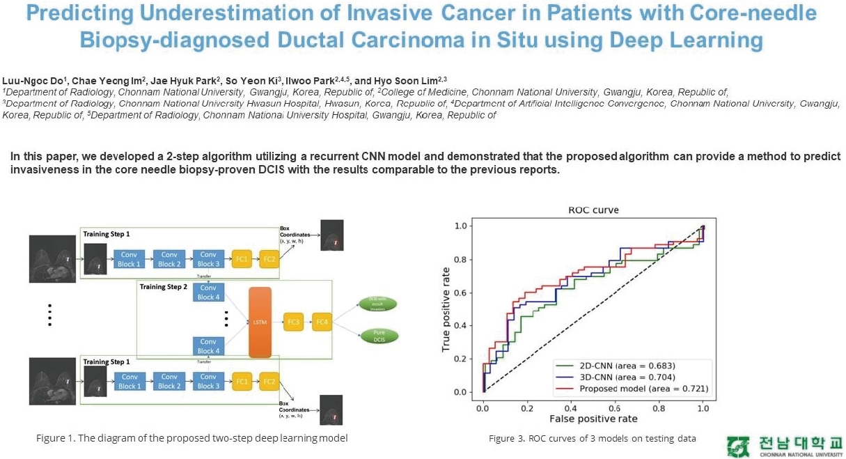
In 2017, the potential applications of AI-generated excitement within the medical profession. Researchers from Stanford University published a research paper in Nature that their convolutional neural network (CNN) algorithm was superior to dermatologists in determining skin cancer classification [1]. Additionally, IBM Watson was introduced in several university hospitals. Professor Geoffrey Hinton, referred to as the father of AI, argued that the radiology department should stop training within 5 years [2]. Simultaneously, it appeared that AI could potentially replace the doctor’s positions and roles at any moment, given these advancements and superiority. As a premedical student, I thought I had to jump into the sea of AI immediately so I would not be superseded or subordinated to future technology. Therefore, I knocked on the “Center for AI in Medical Imaging Research” door at the Chonnam National University Hospital to get involved, and professor Il-woo Park (Department of Radiology, Chonnam National University Medical School) gratefully allowed me to participate in their ongoing breast cancer research. As a result of these efforts, we developed a two-step CNN algorithm that predicts cancer invasiveness in patients with core-needle biopsy-diagnosed ductal carcinoma in situ [3]. Furthermore, he taught and mentored me to publish abstract research papers in the 2021 International Society for Magnetic Resonance in Medicine as co-author of the ‘Predicting underestimation of invasive cancer in patients with core-needle biopsy-diagnosed ductal carcinoma in situ using deep learning’ (Fig. 1).
2) Four years of ‘college of medicine’ period and ‘meningioma’ research
For my first 2 years in the college of medicine, my academic burden made it challenging to participate continuously in AI-driven research. Therefore, I concentrated on studying basic medicine and clinical medicine for 2 years. However, my interest in clinical AI research was still present, so I quenched my thirst for AI research by participating in several research internships like ‘Severance Advanced Clerkship (Artificial Intelligence-Powered Radiology)’ at Yonsei University College of Medicine and “the 29th Clinical Pharmacology Internship” at Seoul National University Hospital during vacation periods. Notably, these medical research experiences motivated me to have the personal goal of graduating from medical school with a research paper that I led and authored.
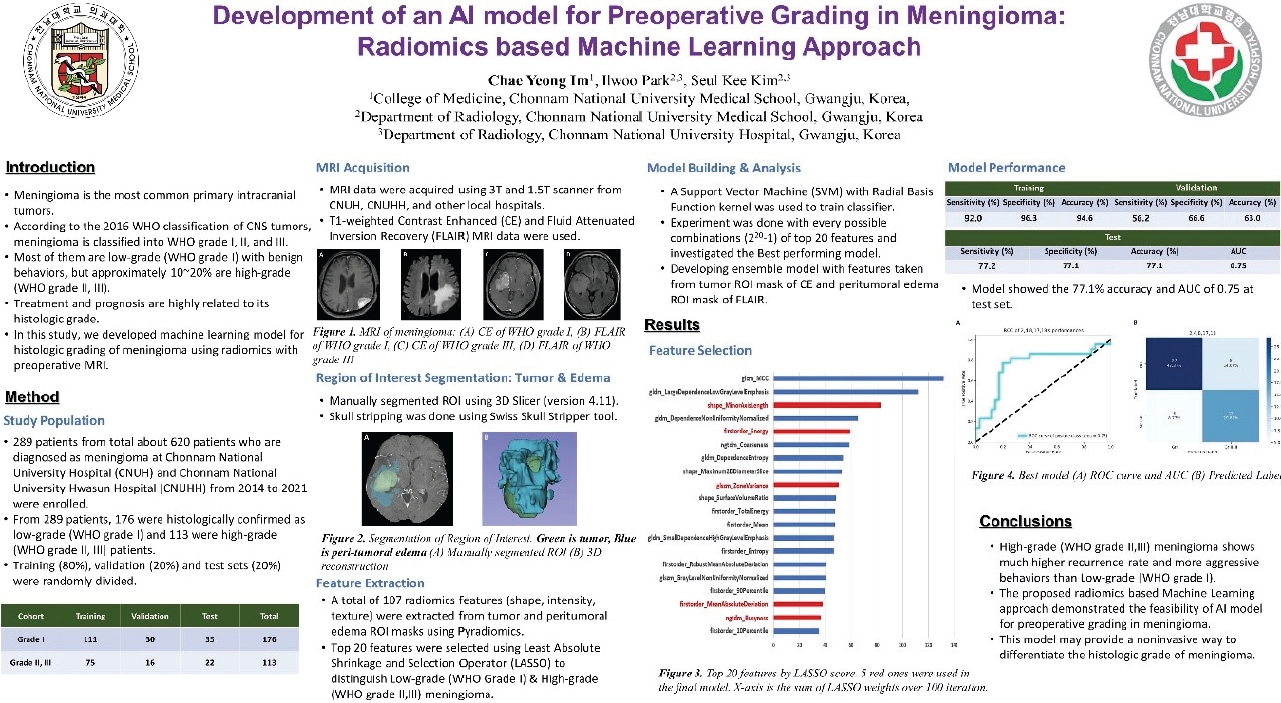
As soon as the winter vacation of my second grade in the college of medicine started, I launched a new research project. For a few months, I learned fundamental knowledge to conduct radiomics experiments. I also had several research meetings with Professor Seul-kee Kim (Department of Radiology, Chonnam National University Medical School) to consolidate our methodology and research direction. Radiomics, a newly developed precision medicine and oncology technology, translates high-dimensional image information into abundant mathematical data by multiple computational algorithms. It provides an objective and quantitative approach to interpreting imaging data, rather than the subjective and qualitative interpretation from relatively limited human visual observation [4]. So, we decided to utilize the radiomics-based machine learning approach to develop our model for preoperative grading in meningioma, the most common central nervous system tumor. Subsequently, during my third year in the college of medicine, I decided to conduct my research in parallel with a clinical clerkship. To be honest, it was much more challenging than I had expected. However, I could not give up and was greatly rewarded during this experience because many professors encouraged and helped me to progress my technical and soft skills. Importantly, I also felt great satisfaction from creating something new that did not exist in the world and pride that I am contributing to improving human health through this research.
November 2021, with the support of professors, I was able to attend the 9th International Congress on Magnetic Resonance Imaging conference and study the latest trends in radiomics and magnetic resonance physics research. At the conference, I had a conversation with my internship advisor, professor Young-han Lee (Department of Radiology, Yonsei University College of Medicine). The Department of Radiology at Severance Hospital was also conducting research similar to what I was investigating at that time. Unlike our method, which only uses radiomics features, their approach combined clinical data with radiomics features [5]. I continued my research until early 2022, when in my fourth year of the college of medicine. I presented our results at the Chonnam National University Medical School Research Conference and won the Best Poster Award (Fig. 2). Currently, this research is expanding, and we plan to progress international validation with foreign university hospitals such as the United States, Türkiye, and Vietnam based on our developed model.
3. Conclusion
While attending medical school in the era of AI, I was able to study and research both medicine and artificial intelligence at the same time. Also, I am greatly proud and satisfied with my medical school years because I could have ramped up my abilities and skills through these education and research experiences. Medicine and engineering both have a common principle in terms of problem-solving. While medicine emphasizes experience and evidence, engineering or AI puts much more importance on developing something superior to experience and creating a brand-new model. The disparity between these two sciences confused me during my research, and learning AI was much more complicated and challenging than I had expected. So, based on my experiences, I would like to propose some educational approaches that can help medical students to learn AI or participate in AI research. First, establishing basic computer programming course for medical AI during a ‘premedical’ period could be helpful. Second, It would be a great chance for medical students to develop their research abilities and skills if clinical research courses are included in the ‘college of medicine’ period.


